In biological research, fluorescence staining is a key technique to reveal the locations and morphology of subcellular structures. However, it is slow, expensive, and harmful to cells. In this paper, we model it as a deep learning task termed subcellular structure prediction (SSP), aiming to predict the 3D fluorescent images of multiple subcellular structures from a 3D transmitted-light image. Unfortunately, due to the limitations of current biotechnology, each image is partially labeled in SSP. Besides, naturally, subcellular structures vary considerably in size, which causes the multi-scale issue of SSP. To overcome these challenges, we propose Re-parameterizing Mixture-of-Diverse-Experts (RepMode), a network that dynamically organizes its parameters with task-aware priors to handle specified single-label prediction tasks. In RepMode, the Mixture-of-Diverse-Experts (MoDE) block is designed to learn the generalized parameters for all tasks, and gating re-parameterization (GatRep) is performed to generate the specialized parameters for each task, by which RepMode can maintain a compact practical topology exactly like a plain network, and meanwhile achieves a powerful theoretical topology. Comprehensive experiments show that RepMode can achieve state-of-the-art overall performance in SSP.
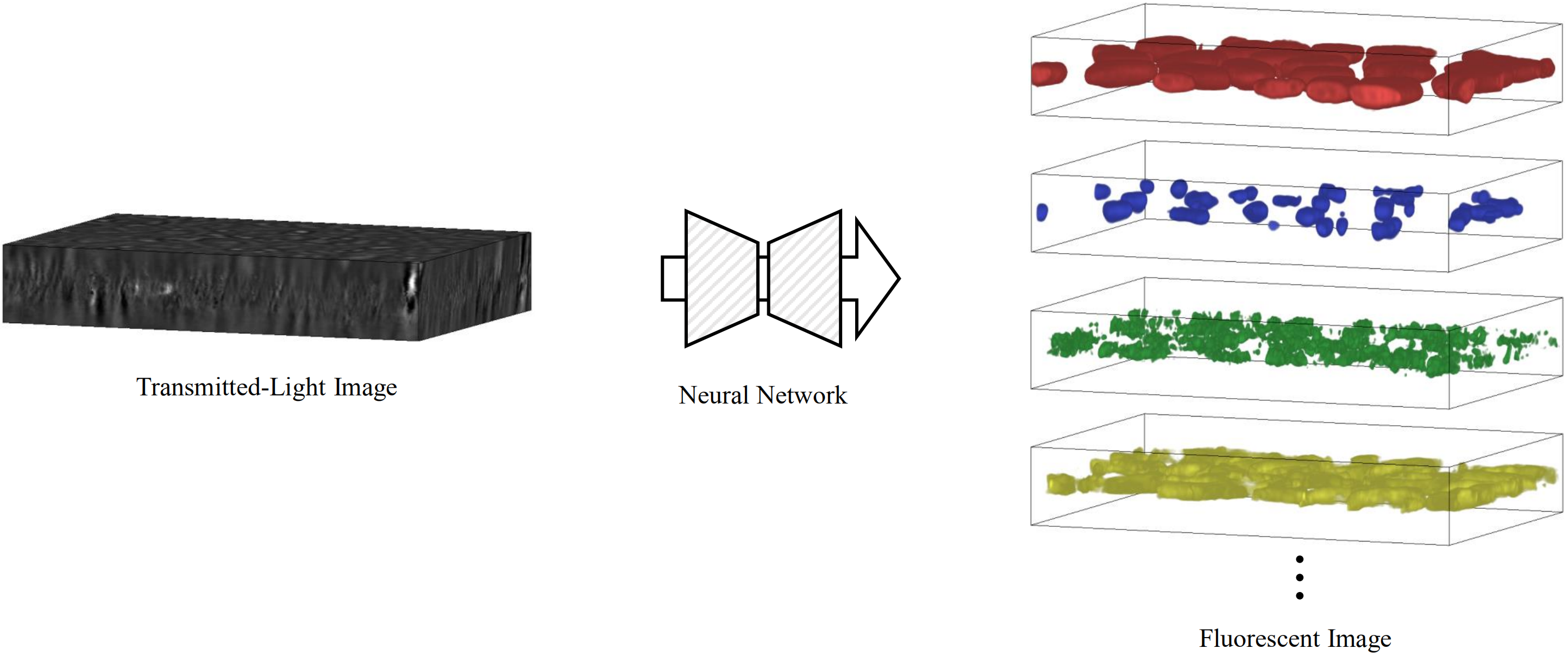
Illustration of subcellular structure prediction (SSP). SSP aims to predict the 3D fluorescent images of multiple subcellular structures from a 3D transmitted-light image. This task faces two challenges, i.e. partial labeling and multi-scale.
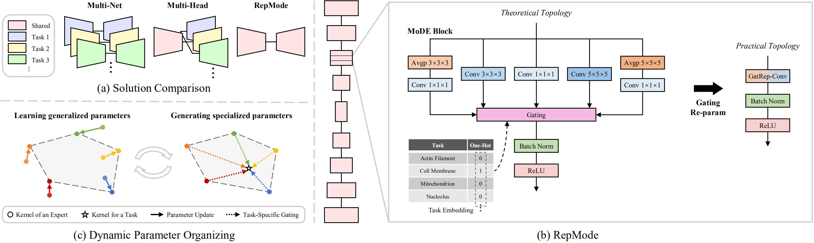
Overview of the proposed method. (a) Comparison of two mainstream solutions (i.e. Multi-Net and Multi-Head) and the proposed method (i.e. RepMode) for SSP. (b) Illustration of our RepMode which includes two key components, i.e. the proposed MoDE block and GatRep. (c) Diagram of how RepMode dynamically organizes its parameters in a MoDE block. Note that the gray region denotes the convex hull decided by the expert kernels, and the convex hull is the area where the task-specific kernels would be situated.

Examples of the prediction results on the test set. These images are from (a) microtubule, (b) actin filament, (c) DNA, (e) tight junction, (f) actomyosin bundle, (g) cell membrane, and (h) endoplasmic reticulum. We compare the predictions of our RepMode with the ones of TGNet (a competitive method). Note that the dotted boxes indicate the major prediction difference.
    @article{zhou2022repmode,
      title  ={RepMode: Learning to Re-parameterize Diverse Experts for Subcellular Structure Prediction},
      author ={Zhou, Donghao and Gu, Chunbin and Xu, Junde and Liu, Furui and Wang, Qiong and Chen, Guangyong and Heng, Pheng-Ann},
      journal={arXiv preprint arXiv:2212.10066},
      year   ={2022}
    }